


向编辑:
结节病的胸淋巴结病通常会显示特征和众所周知的特征,例如对称的肺野马露的分布和非压迫性质。然而,压缩淋巴结病称为疾病的非典型且可能的呈现。若干案例报告和评论先前报道了洛巴寡石或节段支气管,肺动脉分支,高级腔静脉或左侧喉部复发神经的压缩1-4.。据我们所知,从未描述过心脏的压缩。我们在此报告了由血管子淋巴结引起的血管压缩四种患者的临床,形态学和功能特征。根据美国胸部社会(ATS)/欧洲呼吸协会(ERS)/世界结节病和其他肉芽肿疾病(灰)标准进行了诊断188bet官网地址5.。一名患者在诊断(Löfgren综合征)上有一个疾病,最终进化到第二阶段。三名患者有两个阶段的结节病;其中一个展示了特征性皮肤参与,另一种宫颈淋巴结病和可能的肌肉受累。在第三名患者中,疾病仅限于胸部,并在随访期间进化到第四阶段。
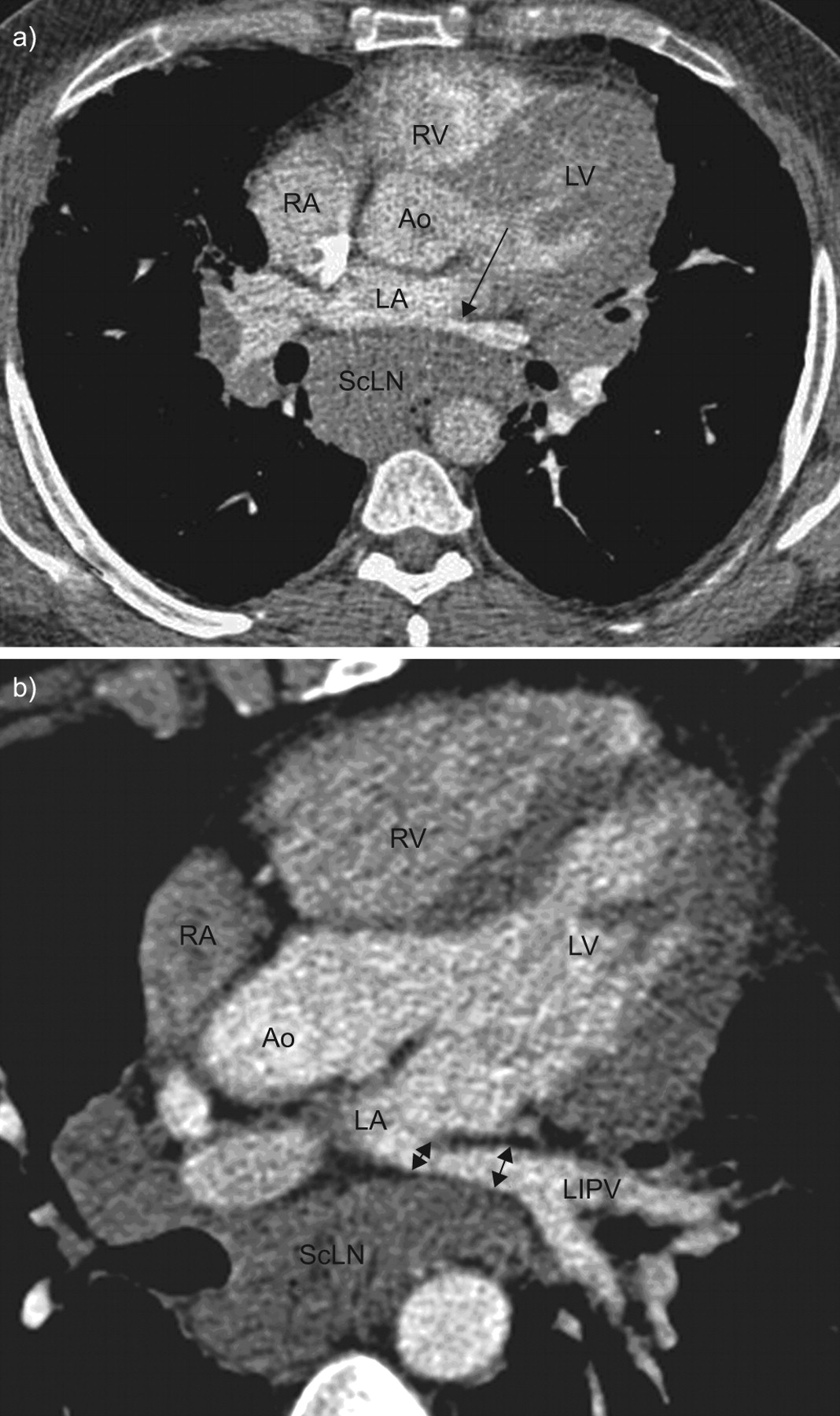
在随访期间执行的计算机断层扫描(CT)扫描变得显而易见。所有患者当时抱怨锻炼的呼吸短促。CT扫描(图1⇓)显示左侧淋巴结肿大的扩大,压缩左心房后壁,使其直接或凸形。对于三名患者,还注意到一种或几种肺静脉的外在压缩,诱导狭窄率达50%(表1⇓)。实质结节病也increased in all patients, but remained moderate for patients one and four. For these two patients, the pulmonary functional tests at rest remained normal, while the two other patients (two and three) exhibited a restrictive pattern with significant alteration of carbon monoxide diffusion. All patients had significant alteration of aerobic capacity (table 1⇓)。Both trans-thoracic and trans-parietal echocardiographies were performed in three patients (one, two and four), and did not reveal pulmonary hypertension but showed left atrial compression. The interatrial septum was deviated towards the right atrium, indicating increased left atrial pressure. The left atrium volume was 15 mL·m-2在患者中(男性参考范围:22±6毫升·m-26.)。休息的卒中量为35毫升·m-2(参考范围:57±14毫升·m-2)休息的心率为90次击败·min-1。The cardiac index at rest was low (3.15 L·min-1·M.-2)。没有潜在的心脏病迹象。心脏磁共振在一种情况下进行,并确认肺静脉的压缩。
a)对阳性病变的阳性增强的计算机断层扫描扫描,显示左侧心房压缩(La)和左下肺静脉(LiPv;箭头)的前静脉狭窄,由扩大的子气态淋巴结(SCLN)引起。b)在同一患者中,倾斜重新格式化图像证实了LIPV(箭头)的狭窄。LV:左心室;ra:右心房;RV:右心室;AO:主动脉。
Characteristics of venoatrial compression and pulmonary function at rest and on exercise
Two of the patients had undergone previous treatment for sarcoidosis, but treatment had been discontinued for at least 1 yr at the time of the left atrial compression diagnosis. Two patients were treated, but decision for therapy was not motivated by the left atrium compression. One patient received corticosteroids (1 mg·kg-1prednisone, gradually tapered) because of the rapid progression of the parenchymal lesions with severe functional impairment. Another patient was treated with infliximab (because of previous severe side-effects with corticosteroids, azathioprine and methotrexate) for incapacitating muscular involvement. In both cases, the size of the enlarged lymph nodes significantly decreased under therapy, and the left atrial compression disappeared (table 1⇑)。
To our knowledge, this is the first description of venoatrial compression in sarcoidosis. Many cases of compression of mediastinal structures by lymph nodes in sarcoidosis have been reported in the literature. Compression of pulmonary arteries by enlarged lymph nodes is considered to be a possible aetiology for pulmonary hypertension in sarcoidosis4.。这种不寻常的压缩性质没有真正的病理生理假设。
当诊断出左心房压缩时,所有病例都被重新评估,无论是替代还是额外的诊断(结节病和癌症或淋巴瘤,结节病和结核病,etc。)。所有的结节病诊断都是根据ATS / ERS / WASOG陈述进行的5.,除了任何患者中的压缩淋巴结外,没有其他非典型特征。新的痰样品对于结核病是阴性的。结核蛋白皮内反应仍然是阴性的。癌症或恶性血症患者没有临床或放射性论证。所有患者均为HIV阴性。我们排除了组织质,因为它在法国的极低普遍存在。最后,我们目前有1-3岁的跟进;两种在皮质类固醇或免疫抑制治疗下已经解决,另外两种也没有进化。没有发生与诊断结节病的诊断发生的新元素。
我们报告的案件是最近的;因此,我们使假设通过顺序化淋巴结左侧压缩可能不是这样一个罕见的实体,并且可能是由于两个原因而受到的。首先,目前尚不清楚左侧心房压缩是否诱导症状,如果是的话,它们将在呼吸系统疾病的环境中是非特异性的。在少数几个评论中的一个解决问题中,d'cruz等等。7.distinguish different degrees in cardiac involvement of mediastinal masses. He describes a mediastinal mass as being “compressive” if it produces haemodynamic effects and symptoms akin to tamponade. On the contrary, a mass labelled as “encroaching on” the heart would narrow or distort it but would not produce haemodynamic effects or clinical symptoms. However, one cannot rule out that a moderate compression or encroachment on the left atrium would produce more subtle haemodynamic alterations and, therefore, symptoms, especially during exercise. Dyspnoea on exercise is reported in several cases of left atrial compression of other causes, such as bronchogenic cyst or hiatus hernia. Though none of our patients presented signs of tamponade or cardiac failure, all of them complained of dyspnoea on exertion and decreased tolerance to exercise. While two of the patients had moderate to severe alteration of carbon monoxide diffusion, the remaining two had a normal or close to normal oxygen saturation and oxygen alveolo-arterial gradient on exercise, as well as a preserved ventilatory reserve. In these patients, the oxygen pulse remained low throughout exercise, suggesting a limitation in the increase of stroke volume on exertion8.那9.由压缩引起的,这损害了左心房的储存功能。在另外两个患者中也发现了低峰氧脉冲,但由于运动的氧气,并且可能的肌肉受累,其解释难以。肺静脉狭窄通常在涉及几个静脉时诱导症状,并且当狭窄> 60%时10.,这不是我们患者的情况。此外,左心房和肺静脉压缩对CT扫描的良好可视化可能需要对比度,这是不常规用于的术语患者。即便如此,左心房的可见压缩可能会被忽视放射科医师和临床医生,以及超声心动图,他们不期望这种异常。在我们两个患者中,在最终诊断之前,在至少一个先前的CT扫描或超声心动图上已经可见了压缩。我们建议仔细注意左心房后壁和大量子地淋巴结患者的肺静脉。
The compression disappeared for the two patients that were treated, which is consistent with many reports about compression of bronchi or pulmonary arteries. It is uncertain whether a left atrial and/or pulmonary vein compression should represent an indication for treatment本身,作为临床相关性和在症状发作中的作用尚不清楚,难以评估。还必须记住,这种压缩可能具有自发性结果。
总之,通过淋巴结病变在结节病中的各种纵隔结构压缩已经被描述为疾病的非典型形式。然而,这是左心房和肺静脉压缩的第一报告。意识到存在这种情况的存在,特别是在显示大亚曲菇淋巴结病的患者中,应增加其诊断,并让我们更好地理解其对运动能力的影响。
兴趣表
None declared.
致谢
T. Le Tutneau,服务D'Explorations FonctionNelles Cardio-Vasculares,HôpitalCediquique,Chru de Lille,Lille Cedex,法国。
- © ERS Journals Ltd


