一个bstract
支气管活检标本可能不充分represent inflammatory cell counts throughout the airway wall. The present study aimed to compare mast cell density in biopsies and airway sections using both stereological and nonstereological methods.
Post mortembiopsies and adjacent transverse sections were obtained from a mean of five proximal airways per case in 10 subjects who had died of nonrespiratory causes. Tryptase-positive mast cells were measured stereologically in 30-μm sections and nonstereologically in 5-μm sections using an optical disector (cells·mm-3) and cell profiles (cells·mm-2), respectively. Reference areas included the inner and total airway wall and to 100 μm below the basement membrane.
Case means, based on four or more biopsy sites, significantly correlated with those on transverse sections for counts over the inner airway wall only, using both stereological and nonstereological methods. Cells·mm-3and cells·mm-2were significantly correlated within all reference areas.
When endobronchial biopsies are obtained from at least four proximal airways per case, inter-subject comparisons of mean mast cell density in the inner airway wall are as well represented by counts on biopsies as they are on transverse sections. This is the case using either three-dimensional, stereological or two-dimensional, nonstereological methods.
Development of the flexible fibreoptic bronchoscope in 19671, followed by the resolution of concerns about the safety of its use2, led to an acceleration in the use of endobronchial biopsy to examine inflammation in the airways of patients with asthma. Since the mid 1980s, the perceived success of endobronchial biopsies in demonstrating submucosal inflammation and the effects of treatment3–16, plus the limitations or complications of indirect methods10,17,18, have helped consolidate bronchial biopsy as the “gold standard” for investigating airway inflammationin vivoin asthma.
尽管如此,仍有人怀疑representativeness of biopsies, with several studies suggesting that “biopsy specimens, being small, may not be “valid” for the airways as a whole”6,10,19.Concerns about the anatomical sampling limitations of only taking biopsies from carinal sites20have added to concerns about the limitations of size, depth and sampling from proximal airways only21.
Studies examining the density of inflammatory cells in the airways in asthma using airway transverse sections frompost mortemtissue sample the bronchial tree more comprehensively than do studies using bronchial biopsies. Since most biopsy studies reporting the density of inflammatory cells have been in mild cases of asthma3,4,6–16,22, comparisons withpost mortemstudies are effectively limited to the small number of such studies that have compared transverse sections from cases of less severe, nonfatal asthma and controls23,24.Results of biopsy studies in mild asthma have not been entirely consistent with either results of thepost mortemstudies using cases of nonfatal asthma, or with each other.
一个nother methodological consideration is whether the density of inflammatory cells in endobronchial biopsy specimens should be assessed by means of traditional two-dimensional (2D) “area profile counts” or three-dimensional (3D) “stereology-based” methods20.一个t the centre of this issue is the question of potential bias. Since the probability of a cell's profile appearing in a 2D section is directly related to the cell's size, shape and orientation relative to the cutting plane, there is an inherent risk that area profile counts are biased in favour of certain cells25,26.However, 3D approaches render the counts free of such biases as they involve counting cells of interest in 3D volumes to give cells·mm-3.In practical terms, this relies on application of the disector principle27.Very few studies of airway inflammation in asthma have adopted stereological approaches26,28–30and of these only three have used a disector to enumerate cell density. No such studies appear to have used the optical disector31–33in thick sections to measure the density of inflammatory cells in actual 3D space.
What is unclear at this stage is the extent and nature of the relationship (if any) between: 1) estimates of inflammatory cell density obtained from endobronchial biopsies at the carinae with inflammatory cell density across the whole airway wall in the vicinity of the biopsy site; and 2) estimates of inflammatory cell density obtained from samples of these tissues with the use of 3D compared with the more commonly used 2D approaches.
The aim of the present study was to compare the density of mast cells in endobronchial biopsies and transverse sections of large airways from corresponding sites, using both stereological 3D and nonstereological 2D techniques.
METHODS
Whole lungs (eight left, two right) were obtained atpost mortemfrom 10 subjects (all male, aged 14–44 yrs) who had died from nonrespiratory causes. Consent for the study of these tissues was obtained from the next of kin and with the approval of the ethics committee of Sir Charles Gairdner Hospital (Nedlands, Western Australia). Medical histories were only available from four subjects and in each case excluded the diagnosis of asthma. Lungs were fixed in inflation with 10% neutral-buffered formalin at a pressure of 25 cmH2O.
Large airways were biopsied under direct vision at the (sub)carinae using standard cupped biopsy forceps. Following each biopsy, a transverse block 2–3 mm long was cut from one of the airway branches immediately adjacent the biopsy site. This procedure was followed progressively from the main lobar bronchi down to fifth or sixth generation airways, taking at least one biopsy and paired transverse block at each level (fig. 1⇓).
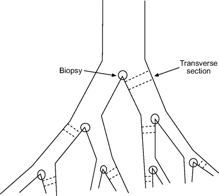
Schematic diagram illustrating biopsy and airway dissection protocol in proximal airways.
For each subject, six biopsies and their paired transverse airway blocks were selected at random from those collected. Each biopsy and transverse airway block was embedded separately in paraffin wax and sections were cut at a thicknesses of both 5 μm and 30 μm. Sections were stained with anti-mast cell tryptase monoclonal antibody AA1 (Dako, Botany, Australia) using the immunoperoxidase technique as previously described34.Sections with preparation artefacts were discarded or replaced.
Cell counting
一个ll cell counts were conducted using the Computer Assisted Stereology Toolbox (CAST) grid® system (Olympus, Ballerup, Denmark), using an oil immersion lens at ×1,000 magnification and numerical aperture setting of 1.35 to minimise the depth of field. Optimal sampling fractions (i.e.the fraction of the area of interest sampled by uniformly spaced, randomly positioned (UR) fields) of the airway wall compartments were based on pilot counts of mast cells on transverse sections. These were conducted on at least seven seven trial sampling fractions of 1, 2, 5, 10, 17 and 25%.
Estimates of 2D mast cell density in 5-μm transverse airway sections
Both the inner airway wall (WAi), from the epithelial basement membrane to the outer border of the airway smooth muscle layer, and the total airway wall (WAt), from the epithelial basement membrane to the outer border of the adventitia35, were delineated on CAST, and, within these reference areas, samples of UR 3,084.3 μm2fields comprising 17% of WAiand 5% of WAtwere generated. These fields were presented sequentially on the high-resolution CAST monitor and positively stained, nucleated cell profiles in focus were counted within a single focal plane with the aid of an unbiased counting frame36.The count was repeated on a second, independent sample of UR fields and the mean of the two independent estimates was calculated.
Estimates of 3D mast cell density in 30-μm transverse airway sections using the optical disector
The optical disector uses a probe that is a predetermined sampling volume within the tissue of interest. The upper and lower borders are preset to avoid the cut edges of the thick tissue sections, and lateral borders are set to allow convenient sampling, depending on the density of objects to be counted. To allow for deformation during tissue processing, the actual thickness of each nominal 30-μm-thick section was estimated optically as the difference in the height of the microscope stage (using microcator readings) from where the tissue first came into focus to where the tissue went out of focus. The average of 10 random points within delineated areas was calculated. Guard areas 4-μm and 5-μm thick were established at the top and bottom of each section respectively (fig. 2⇓), with the balance of the section's mean thickness being set as the height of the optical disector probe. This resulted in an average disector height of 17 μm. Further guard areas 14–18-μm wide were established around the sides of the disector.
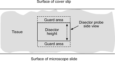
Schematic side view of the optical disector showing the guard areas above and below disector probe (not to scale).
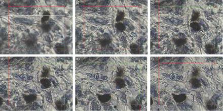
To count positively stained cells appearing in a field, the focal plane with its associated unbiased counting frame was scanned slowly down through the tissue. Cells first coming into focus within the disector height were counted provided that they fell entirely within the counting frame or only touched its acceptance lines and not its forbidden lines (fig. 3⇓).
Histological staining of positively stained lung tissue cells at a depth of a) 5 μm, b) 10 μm, c) 15 μm, d) 20 μm, e) 25 μm, and f) 27 μm. The size of the counting frame (solid white line) is 64 μm. The unbiased counting frame is scanned slowly down through the tissue. A cell is counted (arrow) if it first comes into focus within the active disector depth and lies wholly or partly within the frame without crossing the (red) exclusion lines. The disector is active when inclusion lines turn green at the specified tissue depth.
Within each airway section, the number density (Nv) of mast cells (cells·mm-3) was estimated for WAiand WAtby:
Nv = (109ΣQ)/(a.h. ΣP) (1)
where ΣQ = total number of positively stained cells counted in the sample, ΣP = total number of fields in the sample, a = area of the unbiased counting frame (μm2), and h = disector height (μm).
Estimates of 2D and 3D mast cell density in endobronchial biopsy sections
Independent 17% samples of UR fields were generated over two reference areas; WAi, where definable, and an area to a depth of 100 μm below the basement membrane, similar to previous studies3,6,7,16.2D counts in 5-μm biopsy sections were conducted as for 5-μm transverse sections to give cells·mm-2.3D cell counts in 30-μm biopsy sections were conducted using the optical disector as for 30-μm transverse sections to give cells·mm-3.
Data analysis
Pearson's correlation coefficient was used to test the relationship between mast cell densities in biopsies and mast cell densities in associated airway transverse sections, and the relationship between 2D and 3D counts. Mean r-values, where shown, are Fisher Z weighted means. Otherwise, where appropriate, data are presented as mean±sd.Intraclass correlation coefficients were also used to assess the relationship between comparable counts on biopsies and transverse sections. Differences between comparable biopsy and transverse section counts were assessed by Bland–Altman plots. The effect of the number of biopsies used was also examined by repeating the analyses using data from one to four biopsies. Correlations were with the WAion all transverse sections. Comparisons of results on biopsies and transverse sections were also made between cases grouped by cell counts obtained on transverse sections. An unpaired t-test was used to assess differences between these groups.
RESULTS
Estimates of intra-observer variability for delineation of reference areas were all <1%. On transverse sections, the mean count areas assessed per section were 1.33 mm2and 2.13 mm2for the WAiand WAt, respectively. On biopsies, the mean count areas assessed per section were 0.29 mm2for the WAiand 0.12 mm2to a depth of 100 µm below the basement membrane.
Relationships between mast cell density in endobronchial biopsies and adjacent airway transverse sections within cases
Table 1⇓shows mean intracase correlations between 2D and 3D mast cell densities in individual biopsies, and 2D and 3D mast cell densities in corresponding airway transverse sections taken adjacent to the biopsy site. In general, mean r-values were low, with no consistent relationship between measurements regardless of the airway compartment measured or the type of measurement (2D or 3D) employed.
Intracase correlations of mast cell densities of biopsiesversustransverse sections
Relationships between case means of mast cell density in biopsies and adjacent airway transverse sections
Mean mast cell densities over the WAiin biopsies showed consistent, significant positive correlation with mean mast cell densities over the WAion airway transverse sections (table 2⇓).There was no systematic difference between comparable biopsy counts and transverse section counts for the WAiand differences were within Bland–Altman 95% limits of agreement in >90% of comparisons. This was irrespective of whether cell counts were carried out in 2D or 3D. However, in general, the biopsy counts were not significantly correlated with mean mast cell density over the WAton transverse sections.
Intercase correlations of mean mast cell densities using all biopsiesversusall transverse sections per case
Mean mast cell densities to a depth of 100 μm below the basement membrane in biopsies showed no consistent, significant correlation with mean mast cell densities over the WAion airway transverse sections, and no significant correlation with mean mast cell densities over the WAton airway transverse sections (table 2⇑).
The effect of using one, two, three or four endobronchial biopsies per case
For the WAi, the density of mast cells based on a single endobronchial biopsy did not correlate with the case mean on transverse sections (table 3⇓).一个lthough mean 2D counts on biopsies showed some significant correlations with case means on airway transverse sections when two or three biopsies were used, correlation coefficients were not consistently significant for both 2D and 3D counts unless at least four biopsies were used to determine the case mean.
Intercase correlations of mast cell densities within the inner airway wall(WAi)#of 1–4 biopsiesversustransverse sections
Relationship between mean 2D and mean 3D mast cell densities
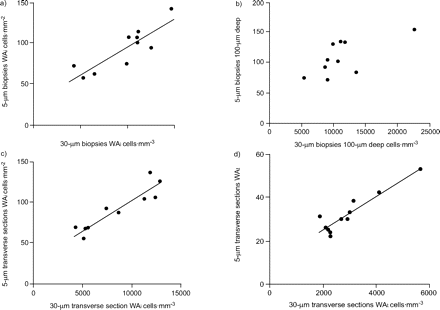
In biopsies, mean 2D mast cell density was significantly correlated with mean 3D mast cell density for counts over both the WAi(fig. 4a⇓) and to a depth of 100 μm below the basement membrane (fig. 4b⇓).In transverse sections, mean 2D mast cell density was significantly correlated with mean 3D mast cell density for counts over both the WAi(fig. 4c⇓) and over the WAt(fig. 4d⇓).In general, 2D and 3D counts on airway transverse sections were more strongly correlated than 2D and 3D counts on biopsies.
Mean two-dimensional mast cell density of 5-μm biopsies (a, b) and transverse sections (c, d; cells·mm-2)versusmean three-dimensional mast cell density of 30-μm biopsies (a, b) and transverse sections (c, d; cells·mm-3).WAi: inner airway wall; WAt: total airway wall. a) r = 0.86, p<0.005; b) r = 0.68, p<0.05; c) r = 0.92, p<0.0005; d) r = 0.93, p<0.0005.
Relative variability of 2D and 3D mast cell densities on endobronchial biopsies and airway transverse sections
Mean coefficients of variation (CV) in mast cell density were similar for 2D counts and 3D counts (32 and 37%, respectively), but were significantly smaller for counts on transverse sections than for counts on biopsies (30 and 39%, respectively). On transverse sections, mean CV was smaller for counts over the WAi(23%) than over the WAt(39%). On biopsies, mean CV was similar for counts over the WAi(39%) and to depth of 100 μm below the basement membrane (39%).
Comparisons between case groups
The usefulness of biopsies to detect differences between case groups was examined by comparing cases grouped according to mast cell densities in the WAion all transverse sections. Both 2D and 3D mast cell counts over the WAion biopsies showed similar significant differences between the groups, as did counts over the WAion transverse sections (table 4⇓).However, biopsy counts confined to a depth of 100 μm below the basement membrane did not show significant differences between the two groups.
Comparison of grouped case data using mean mast cell densities in transverse sections and in biopsies#
DISCUSSION
Endobronchial biopsy has been widely used as a research tool to assess airway inflammation in asthma3,4,6–16,22.Its use as a tool in clinical research has recently become the subject of review20.The present study attempted to address the issue of whether endobronchial biopsy specimens provide adequate representation of the distribution of inflammatory cells in the airway wall as a whole. The current study reported a direct comparison of the density of inflammatory cells in endobronchial biopsies and airway transverse sections from the same proximal airways, compared 2D cell profile counts with 3D stereological counts of airway inflammatory cells, and used an optical disector to enumerate the density of airway inflammatory cells in actual 3D space. In addition, it addressed empirically the issue of the minimum number of endobronchial biopsies required to adequately represent the density of inflammatory cells in proximal airways.
The present study found that, for a given case, any single biopsy is unlikely to be representative of mast cell density across the airway wall in the vicinity of the biopsy site or in the proximal airways generally. However, four or more biopsies from different sites can be used to generate a case mean which is likely to be representative of mast cell density across the WAiin proximal airways for the purpose of comparing cases. Both 3D stereological and 2D nonstereological methods of assessing mast cell density in proximal airways resulted in similar conclusions. There was little evidence that intercase comparisons of mast cell density in endobronchial biopsies parallel intercase comparisons of mast cell density across the WAtin proximal airways, or that measurements of mast cell density confined to a depth of ≤100 μm below the basement membrane on endobronchial biopsies can adequately represent mast cell density across the remainder of the airway wall in proximal airways.
The use of a single endobronchial biopsy
一个number of biopsy studies of airway inflammation appear to have relied upon the use of a single biopsy specimen8,13–15.在目前的研究中,意味着intracase相关性s between mast cell density in biopsy sections and adjacent airway transverse sections were very low. This suggests that within a case, any given endobronchial biopsy from a (sub)carina is unlikely to reflect mast cell density across the airway wall generally in the vicinity of the biopsy site. Such a finding is not unexpected given previously observed intrasubject variations in the density of inflammatory cells within sections37and between sections8,38from the same biopsy site, and between biopsy-sized segments within an airway section39.
此外,肥大细胞密度之间的相关性in one randomly chosen biopsy per case, with the corresponding case mean of mast cell density in airway transverse sections, did not suggest that a single biopsy is likely to adequately reflect mast cell density in the remainder of the bronchial tree for the purposes of intercase comparison. In the current study, single biopsies reflected, at best, ∼30% of the variance in mean mast cell density in the WAion transverse sections from proximal airways.
These results are consistent with the finding in a recent study by Gambleet al.40that sampling more than one airway level is preferable in order to discriminate case groups in chronic obstructive pulmonary disease on the basis of inflammatory cell density in bronchial biopsies.
Case means from multiple endobronchial biopsies
WAi
The inherent difficulties and potential risks of obtaining multiple biopsiesin vivoand the problem of occasional artefactual damage tend to restrict the number of biopsies used in research settings. Biopsy studies of airway inflammation have generally used only two or three biopsies per case to enumerate a particular cell type3,4,6–10,12–16, and have often relied on 2D cell profile methods, expressing density as cells per area4,8–12,14–16.
The results of the present study indicate a progressive strengthening of the relationship between mast cell counts in the WAion biopsies and transverse sections as more biopsies are used. Single biopsies reflected <30% of the variance in mast cell density in all transverse sections. However, this proportion increased as more biopsies were used to generate a case mean until, using four or more, it was >50%, ranging upwards to ∼70%. Single biopsies were not significantly correlated with transverse sections at all, while the only significant correlation between two biopsies and all transverse sections was confined to 2D counts in both, which are potentially biased. In contrast, 2D case means obtained by using three biopsies were significantly correlated not only with 2D counts on transverse sections, but also with 3D counts, which arguably give the best unbiased estimates of cell density for the proximal airways. However, the most consistent results were obtained using four or more biopsies, since mean mast cell densities in the WAion biopsies and transverse sections were significantly correlated regardless of whether 2D or 3D counts were used. These observations are supported by intraclass correlations, which tend to accentuate the differences between using four or more biopsies and fewer than four biopsies.
In essence, the current findings suggest that mast cell density across the WAiin proximal airways is well represented by the mean of four or more biopsies. The mean of two or three biopsies may be adequate, but there is more uncertainty associated with the use of three biopsies than with four or more, and much more uncertainty with two. A single biopsy seems unsatisfactory.
WAt
However, the results did not indicate that intercase comparisons of mean mast cell density over the WAion biopsies parallel intercase comparisons of mast cell density over the WAton transverse sections to a similar degree. Even when using all available biopsies, the resulting case mean generally reflected only 30–40% of the variance in mean mast cell density over the WAton transverse sections. In part, this would have been due to greater heterogeneity in the distribution of mast cells over the WAtin proximal airways when compared with the WAi23, as a result of differences between these compartments in the presence or distribution of structural elements, such as smooth muscle, cartilage plates, submucosal mucous glands and blood vessels.
Cell counts to a depth of 100 μm below the basement membrane on endobronchial biopsies
一个number of biopsy studies reporting the density of airway inflammatory cells, including mast cells, have based their results explicitly on cell counts taken to a depth of approximately ≤100 μm below the basement membrane3,6,7,16.Establishing such a reference area in biopsies is almost always possible as there is usually sufficient countable tissue present, and may be largely a matter of convenience since a 10×10 eyepiece graticule represents a width of 100 μm when using a ×100 objective lens in light microscopy.
The results of the present study did not indicate that intercase comparisons of mean mast cell density based on such a reference area in biopsies parallel intercase comparisons of mast cell density over the WAigenerally, or the WAtgenerally, in proximal airways. Mean mast cell density to a depth of 100 μm below the basement membrane on biopsies reflected, on average, only ∼32% of the variance in mean mast cell density in the WAion transverse sections, and ∼11% of the variance in the WAt.Comparison of mean mast cell densities based on this reference area in biopsies did not reflect the significant differences between subjects grouped by mean cell density in the WAi.
Mast cells are widely distributed across the WAi, including the smooth muscle layer23.一个lthough some authors have implicitly endorsed cell counts in biopsies to a depth of 100–125 μm below the basement membrane, mostly for a variety of other inflammatory cells40–42, a potential problem with using a narrow band of tissue of fixed absolute width when enumerating mast cells is that it will represent a variable fraction of the width of the WAiat the biopsy site, generally a smaller fraction for larger airways. It may include the smooth muscle layer in the smaller proximal airways, but not in the larger ones. Therefore, it would seem advisable, when counting mast cells within the WAi, to extend the count area to the outer border of the smooth muscle layer. However, while the probability of obtaining “assessable” smooth muscle in an endobronchial biopsy specimen may be ∼70% in segmental airways, it may be much less in the lobar bronchi43,44.
2Dversus3D counts
Some theoretical and practical considerations
Differences of opinion exist among investigators regarding the relative merits of using 3D stereological methods compared with 2D nonstereological methods to assess the density of inflammatory cells in airway tissue20,45.The two methods produce entirely different quantities. The use of uniform randomised sampling and 3D counting in thick histological sections with an optical disector enables a direct and unbiased estimate of the number density of cells within tissues, expressed as cells per unit volume. Conversely, 2D counts provide an indirect indication of the number density of cells, based on cell profiles per unit area. Moreover, as demonstrated recently in a study by Fehrenbachet al.26, comparing 2D and 3D quantification of inflammatory cells in bronchial biopsies, such counts may be subject to bias since the probability of a cell’s profile being included in a thin histological section is proportional to cell height, shape and orientation perpendicular to the plane of the section.
Counting cells in 3D with an optical disector requires the use of relatively sophisticated, specialised and expensive apparatus, such as the CASTgrid® system (Olympus), which was used in the present study. This consisted of: 1) an upright light microscope fitted with a video camera, a microcator and a motorised stage driven by a joystick; 2) an XYZ reader; 3) a computer with a high-resolution monitor; and 4) associated CASTgrid® software. The actual disector method of counting cells with this equipment is, however, relatively simple (fig. 3⇑).当前作者发现计数单元的公关ofiles in 2D was simpler, faster and cheaper than counting in 3D with an optical disector, and by requiring only single thin sections, was more economical in the use of limited biopsy tissue. Thin sections were also less prone to tissue disruption in processing.
Nevertheless, it was found that some features of a stereology workstation could be used to good effect in counting inflammatory cell profiles in 2D, as well as counting in 3D. These included: 1) computerised delineation of the reference area; 2) uniform random sampling of the reference area using the meander sampling facility; 3) counting cell profiles with the aid of an unbiased counting frame; 4) viewing serially presented high-power fields (×1,000 plus) on a high-resolution monitor; 5) use of the navigator facility for easy visualisation of field location within the reference area; 6) marking counted cells with customised icons; and 7) automatic logging of relevant count data.
Use of the above techniques in 2D counts does not rule out the potential bias that is inherent in such counts. However, delineation of reference areas at low magnification, followed by UR sampling and counting with an unbiased counting frame and associated inclusion rules at high magnification, may rule out other subtle biases pertaining to identification and inclusion of cells when relying on more traditional manual “randomisation” of fields and the use of an eyepiece graticule.
Current findings
一个s would be expected, 2D and 3D methods produced very different sets of numbers in the present study, with the order of magnitude of mean mast cell densities being mostly 101cells·mm-2and 103cells·mm-3for 2D and 3D counts, respectively. Nevertheless, corresponding 2D and 3D case means displayed very strong positive correlations. This relationship was generally tighter in airway transverse sections where mean 2D counts reflected, on average, ∼86% of the variance in mean 3D counts in both the WAiand WAt.In biopsies, mean 2D counts across the WAireflected ∼74% of the variance in comparative 3D counts. In counts to a depth of 100 μm below the basement membrane, this proportion was ∼45%.
When all available biopsies were used, both 2D and 3D counts resulted in similar comparisons between case means on biopsies and transverse sections. In these comparisons, the strongest correlations occurred between counts across the WAi, where 2D and 3D counts on biopsies were each closely related to both 2D and 3D counts on transverse sections. However, when only three biopsies were used to produce a case mean, the relationship persisted for 2D counts on biopsies, but not for 3D counts.
It is also notable that when cases were grouped according to means of mast cell density across the WAion transverse sections, 2D and 3D counts resulted in the same grouping, and that comparisons between these groups were preserved in both 2D and 3D counts across the WAion biopsies.
When comparing 2D with 3D counts from the same reference area on histological sections, the ratio of (cells·mm-2)/(cells·mm-3) provides a rough estimate of the mean cell height, perpendicular to the plane of the section25.一个veraging this ratio for different count areas across cases gave an estimated mean cell “height” for mast cells of ∼11 μm in biopsies and ∼12 μm in transverse sections. These figures are consistent with reported values for the diameters of mast cells in human lung tissue fixed in formalin46.Thus, the physical height of the relatively rounded, discrete mast cell accounts for the difference in order of magnitude between 2D and 3D mast cell densities.
Generalisability of the findings
The present study focused on different methods for estimating inflammatory cell density in proximal airways. Only mast cells were used for comparing these methods. Mast cells were chosen for initial study for four reasons: 1) they are potent and constitutive inflammatory cells long associated with asthma and now the subject of renewed interest47–49; 2) they have been shown to stain clearly with the marker for mast cell tryptase employed in the formalin fixed tissues used in the current authors’ laboratory; 3) their counts have been found to be highly reproducible; and 4) in proximal airways, they are distributed relatively widely across the airway wall, including the smooth muscle layer and mucous glands23,44, although their absolute density and level of activation may vary in different compartments and disease states23.
Unlike many other studies reporting mast cell density in proximal airways, the present study did not compare subjects with known disease (e.g.asthma) and control subjects. It could be argued that cases grouped on the basis of disease are likely to have a much greater range of data than that observed in the current study, and it is therefore possible that fewer biopsies than the average of four suggested here would be sufficient to discriminate case groups. Due to differences in methods of measuring and/or reporting mast cell densities, it is difficult to compare the data from the current study directly with the data from other studies examining mast cell density in proximal airways in disease states. However, in the few instances where a comparison seems possible, the order of magnitude of mast cell densities is similar, while the spread of data values in the present study appears slightly greater than in some studies12, and slightly less than in others8,23.
The current results were based on data from only 10 subjects. If it had been possible to use lung tissue from much larger numbers of subjects, it is more likely that case means based on fewer than four biopsies would have been correlated significantly with case means over the WAiin all transverse sections, adding weight to the argument that two or three biopsies could be sufficiently representative of the WAigenerally in such circumstances. However, in biopsy studies of airway inflammation in asthma, the size of case groups representing disease states is typically of the order of 10 subjects3,4,7,8,11,12,15,16,19, rarely much more6, and in the case of control subjects, often much less4,7–9,11,12.Furthermore, increasing the number of subjects is unlikely to substantially alter r values, so that while four or more biopsies will probably reflect >50% of the variance over the WAigenerally, fewer than four biopsies will probably reflect much less.
The results obtained will not necessarily be generalisable to other inflammatory cells, since their distributions vary from that of the mast cell. For example, the density of neutrophils and activated eosinophils in the submucosa is not only much less than that of mast cells6,8, but their density relative to that of mast cells appears to vary according to sub-compartments, such as mucous glands34and airway smooth muscle50.Lymphocyte numbers are much higher in the airway wall6,24and their density is subject to much greater variability51.Furthermore, the mast cell appears to be by far the predominant inflammatory cell within the layer of smooth muscle52.Therefore, when counting other inflammatory cells, the inclusion or exclusion of smooth muscle in the measurement area may have a significant effect on overall cell density.
Finally, the density and distribution of inflammatory cells in the small peripheral airways may differ from that observed in the larger proximal airways23,24.Therefore, the relationship between inflammatory cell density in endobronchial biopsies and that in the remainder of the bronchial tree and throughout the lung requires further study.
一个cknowledgments
The authors would like to thank H.J. Gundersen, Stereological Research Laboratory (University of Aarhus, Denmark), and A. Baddeley, School of Mathematics and Statistics (University of Western Australia) for their helpful comments and suggestions.
- ReceivedMarch 15, 2006.
- 一个cceptedJune 30, 2006.
- © ERS Journals Ltd
References